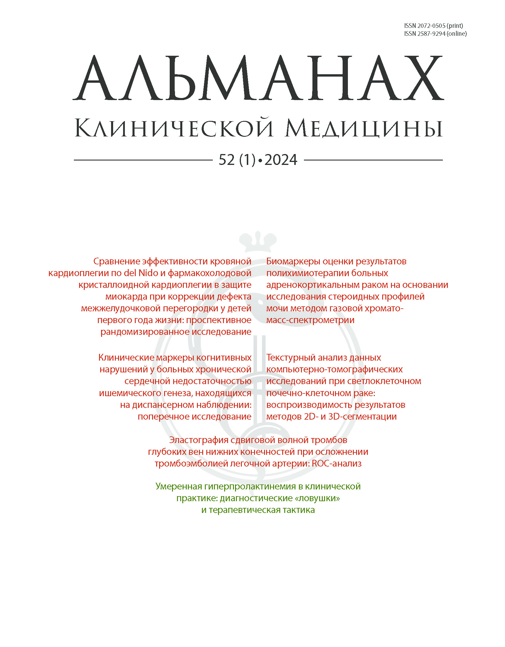
Vol 52, No 1 (2024)
ARTICLES
Del Nido versus cold crystalloid cardioplegia for myocardial protection during ventricular septal defect repair in children under one year of age: a prospective randomized trial
Abstract
Rationale: The choice of strategy for myocardial protection during procedures with cardiopulmonary bypass and cardioplegic arrest in children is not regulated by clinical guidelines due to insufficient data from clinical studies. The issue of methods to assess myocardial injury remains unresolved.
Aim: To assess the frequency and specifics of the development of intraoperative myocardial injury syndrome in children of the first year of life with ventricular septal defect depending on the strategy for cardioplegia.
Materials and methods: In a single center, prospective, randomized controlled trial we compared two cardioplegia strategies during surgical closure of ventricular septal defect in infants aged from 1 to 12 months: del Nido blood cardioplegia (n = 102) and cold crystalloid cardioplegia with Custodiol solution (n = 102). The primary endpoint was a persistent over 10-fold increase above the upper limit of the normal in the plasma concentration of high-sensitivity troponin I at 6 hours after surgery persisting after 24 hours. The secondary combined endpoint was myocardial damage verified by persistent increase in troponin I level more than 10-fold above the upper limit of the normal, persisting at 6 and 24 hours, accompanied by new pathological Q waves, acute complete left bundle branch block, abnormalities of the end part of the ventricular complex on the electrocardiography (ST segment elevation > 1 mm or ST depression of > 1 mm in more than 2 adjacent leads), and a decrease in the global longitudinal strain of the left ventricle by 50% from the initial value at 6 hours after surgery.
Results: In 53/204 (26%) patients, the increase in troponin I persisted at 24 hours after the surgery and was associated with electrocardiography abnormalities, changes in the parameters of left ventricle longitudinal mechanics, and in some cases required greater inotropic support. By the end of the 1st postoperative 24 hours, the longitudinal strain of the left ventricle showed more negative changes over time in the Custodiol group compared to that in the del Nido group (-10 [-14.1; -6.27] versus -14.8 [- 16.5; -10]%; p < 0.0001). The same was true for the left ventricle global strain rate (-0.71 [-0.9; -0.52] s-1 in the del Nido group and -0.57 [-0.760; - 0.44] s-1 in the Custodiol group; p = 0.0049). The primary endpoint was achieved by 21 (20.6%) and 55 (53.9%) patients in the del Nido and Custodiol groups, respectively (p = 0.032). The combined endpoint in the Custodiol group was achieved by 34 (33.3%) versus 19 (18.6%) patients in the del Nido group (p = 0.049, χ2 = 3.875, DF = 1, φ = 0.191).
Conclusion: Del Nido blood cardioplegia compared to cold crystalloid cardioplegia with Custodiol has advantages in terms of preventing intraoperative myocardial damage and minimizing its severity. When assessing myocardial damage, such indicators as left ventricle global longitudinal strain and left ventricle global strain rate are informative, along with an increase in the troponin I level.
 1-9
1-9


Shear wave elastography values of thrombus in patients with lower extremity deep vein thrombosis for pulmonary embolism detection: the ROC analysis
Abstract
Rationale: Thrombosis of the iliac (IV) and femoral veins (FV) is one of the most common causes of pulmonary embolism (PE). Modern ultrasound scanners are equipped with the technology of shear wave elastography, which gives a quantitative assessment of thrombus stiffness by Young's modulus reconstruction. However, the lack of convincing data on the role of thrombus stiffness for clinical manifestations of PE hinders the active use of shear wave elastography to diagnose the risk of embolism.
Aim: To determine the threshold values of the venous thrombus Young’s modulus for deep venous thrombosis (DVT) of the lower extremities complicated by massive PE and/or PE with acute cor pulmonale (ACP).
Materials and methods: This was a single center cross-sectional study in 101 patients who were hospitalized with the diagnosis of acute (duration of less than 2 weeks) or subacute (from 2 weeks to 3 months) IV and FV thrombosis. Doppler ultrasound of the lower extremity veins and echocardiography were done in all patients. Forty eight patients with clinical signs of PE had chest computed tomography. The venous thrombus stiffness was assessed by shear wave elastography with the Young's modulus reconstruction. We performed the ROC analysis for mean values of the Young's modulus for proximal segments of IV and FV thrombi in patients with DVT and massive PE and ACP.
Results: PE was diagnosed in 40.6% (26/64) of the patients hospitalized with acute DVT and in 54.1% (20/37) of those with subacute DVT. Echocardiographic signs of ACP in massive PE were found in 47.4% (9/19) of the patients, in submassive and minor PE in 55.6% (15/27). In DVT complicated with PE, the ROC analysis of the shear wave elastography results gave the following threshold values of the mean Young’s modulus for the proximal thrombus segment: for acute IV thrombosis + PE and ACP, ≤ 16.7 kPa (AUC 0.714, sensitivity 100%, specificity 42.1%), in subacute IV thrombosis + PE and APC, ≤ 23.7 kPa (0.939, 100 and 90.9%, respectively), in acute FV thrombosis + massive PE, ≥ 9.5 kPa (0.706, 100 and 50%, respectively), in subacute FV thrombosis + massive PE, ≥ 24.4 kPa (0.550, 60.0 and 68.8%, respectively).
Conclusion: Shear wave elastography of deep vein thrombi of the lower extremities makes it possible to identify patients with PE and ACP during acute and subacute IV thrombosis and to determine massive PE in acute FV thrombosis.
 10-16
10-16


Biomarkers for assessment of the polychemotherapy results in patients with adrenocortical cancer based on gas chromatography-mass spectrometry studies of urine steroid profiles
Abstract
Background: The effectiveness of polychemotherapy (PCT) for adrenocortical cancer (ACC) is assessed by imaging tests with the RECIST 1.1 criteria. However, the presence of subclinical tumor foci does not allow for an objective measurement of the true tumor burden. As shown previously, postoperative assessment of the steroid metabolome by gas chromatography-mass spectrometry (GCMS) in ACC patients makes it possible to identify early signs of adrenal steroidogenesis abnormalities and of the recurrence of adrenocortical carcinoma.
Aim: To identify biomarkers of response to PCT by GCMS study of the urine steroid profile in ACC patients after surgical resection of the tumor.
Materials and methods: Urine steroid profiles were studied by GCMS (Shimadzu GCMS-TQ8050 gas chromatography-mass spectrometer) in 30 ACC patients (stages II, III and IV) after surgery and first line (combination of etoposide, doxorubicin and cisplatin with daily mitotane) and second line (gemcitabine combined with capecitabine and mitotane) PCT. The control group included 25 patients with hormonally inactive adenomas.
Results: The response to PCT according to RECIST 1.1 criteria was obtained in 23 patients (Group 1, responders) and in 7 patients ACC progressed under PCT (Group 2, non-responders). In the responders, the urinary excretion of etiocholanolone, pregnanediol and pregnanetriol was lower than in the control group. The non-responders had higher urinary excretion of androgens, progestogens and tetrahydro-11-deoxycortisol (THS), compared to the responders and the control group. The patients with ACC progression under PCT had an increase in 3β,16,20-pregnenetriol (3β,16,20-dP3) levels and a decrease of the 3α,16,20-dP3/3β,16,20-dP3 ratio, compared to those in the PCT responders. The threshold values for urinary excretion of dehydroepiandrosterone (DHEA, ≤ 469 mcg/24h; AUC = 1.0), THS (≤ 223 mcg/24h; AUC = 1.0), and 3β,16,20-dP3 (≤ 130 mcg/24h; AUC = 0.986), as well as the 3α,16,20-dP3/3β,16,20-dP3 ratio (≥ 2.13; AUC = 1.0) had 100% sensitivity and specificity for the assessment of the PCT effectiveness.
Conclusion: Different urine steroid profiles were obtained by GCMS in the ACC patients after PCT with and without treatment response. The 100% sensitivity and specificity of the threshold values for urinary excretion of DHEA, THS, 3β,16,20-dP3 and the 3α,16,20-dP3/3β,16,20-dP3 ratio for the assessment of PCT results indicate the potential to use these parameters as biomarkers of response or progression of the disease in the monitoring of PCT effects in ACC patients.
 17-24
17-24


The texture analysis of computed tomography studies in clear cell renal cell carcinoma: reproducibility of 2D and 3D segmentation
Abstract
Background: Differentiation of tumor grade at the preoperative stage is of utmost importance for the modification of the treatment strategy and the extent of operation. However, the routine analysis of computed tomography (CT) data in clear cell renal cell carcinoma (ccRCC) does not allow for reliable determination of the tumor grade.
Aim: To assess the reproducibility of the results of 2D and 3D segmentation of a kidney tumor in the cortico-medullary and nephrographic phases of CT studies, as well as the reproducibility of the first order texture parameters for 2D and 3D tumor segmentation in patients with verified ccRCC.
Materials and methods: This retrospective study included the CT data of 50 patients with morphologically verified ccRCC obtained before their surgical treatment. The first patient group included the patients with the renal tumor size in the axial plane of ≥ 4 cm (28 patients, 29 CT studies), and the second patient group included those with the renal tumor size in axial plane of < 4 cm (22 patients, 23 CT studies). Two radiologists independently performed segmentation of the renal tumor in the cortico-medullary and nephrographic phases of CT procedures done under a standard protocol with the bolus intravenous contrast enhancement. A two-dimensional region of interest (2D ROI) was selected by the investigators on a subjectively selected axial slice, where the tumor had the largest size. When forming a three-dimensional region of interest (3D ROI), the entire tumor volume was segmented. Next, the statistical analysis of the segmentation results and the results of calculation of the first order texture indices was performed with calculation of the intra-class correlation coefficient (ICC) to assess the strength of the data correlation. The ICC of ≥ 0.75 demonstrated the reproducibility of the segmentation results and the first order texture indices.
Results: The 3D segmentation method for ccRCC demonstrated the best ROI reproducibility results, regardless of the tumor size and the phase of contrast enhancement, with the ICC values of 0.961 (95% confidence interval: 0.946–0.971) for the cortico-medullary phase and 0.969 (95% CI: 0.958–0.977) for the nephrographic phase. The 2D tumor segmentation method showed unsatisfactory ROI reproducibility, with the ICC values of ≤ 0.058; however, the unsatisfactory reproducibility of the segmentation results in the patients with ccRCC tumor size of ≥ 4 cm did not significantly affect the reproducibility of the Entropy and Energy texture indices (good to excellent correlation). With the 3D segmentation of ccRCC, most first-order texture metrics were reproducible, with the exception of the Kurtosis parameter. The Entropy and Energy scores in both patient groups demonstrated a high degree of reproducibility. In the 2D tumor segmentation, high reproducibility of the first order texture metrics was obtained for the Entropy and Energy indices.
Conclusion: The 3D segmentation of the CT data for ccRCC has high reproducibility, the most first-order textural features were excellently reproducible when segmentations were performed in 3D. The 2D CT data segmentation method for ccRCC demonstrated low reproducibility; however, some of the first order texture indices were reproducible. Both segmentation methods can be used for the texture analysis of CT images.
 25-34
25-34


Clinical markers of cognitive impairment in patients with chronic heart failure of ischemic origin during out-patient regular follow-up: a cross-sectional study
Abstract
Background: Cognitive impairment (CI) is present in 25–50% of chronic heart failure (CHF) patients. Doctors who monitor patients with cardiovascular disorders do not have clearly set criteria for their referral to a neurologist in case of suspected CI. Therefore, CHF patients do not receive treatment for CI on time.
Aim: To identify significant clinical markers of CI in patients with CHF of ischemic origin.
Materials and methods: This cross-sectional cohort study included 134 patients with CHF of ischemic origin (mean age 63.36 ± 10.63 years; men, 76.12%), who were regularly monitored in a municipal polyclinic. All patients were tested for CI with the Montreal Cognitive Assessment Scale (MoCA); basic hemodynamic parameters, lipid profile, brain natriuretic peptide (NT-proBNP) were assessed, and triglyceride-glucose index (TyG) and body mass index (BMI) were calculated. Cardio-ankle vascular index (CAVI) was measured, echocardiography and a 6-minute walk test (SMWТ) were performed and past history of CHF, arterial hypertension (AH) and diabetes mellitus (DM) was collected.
Results: CI (MoCA score ≤ 25) was detected in 85 (63.43%) outpatients with CHF of ischemic origin; the group without CI (MoCA score > 26) included 49 (36.67%) patients. There were significant correlations between MoCA and CAVI scores (partial correlation coefficient, r = -0.802, p < 0.001; adjusted squared multiple correlation coefficient (adj. R2) = 0.881, p < 0.001), MoCA and TyG (r = -0.357, p = 0.029; adj. R2 = 0.363, p < 0.001), MoCA and SMWТ (r = -0.211, p = 0.037; adj. R2 = 0.696, p < 0.001). The multivariate test for significance of planned comparisons between CAVI and MoCA scores (Wilks' lambda) was 0.005 (F = 4.74; p < 0.001).
Conclusion: CAVI, TyG and SMWТ values are the clinical markers of CI in patients with CHF of ischemic origin. There is a direct association between increased CAVI and the presence of CI, regardless of age, lipid metabolism parameters, structural and functional heart parameters, CHF duration, AH and DM. Identification of these markers could be an indication for an in-depth assessment of CHF patients by a neurologist.
 35-44
35-44


REVIEW ARTICLE
Mild hyperprolactinemia in clinical practice: the diagnostic “traps” and treatment strategy
Abstract
Real world clinical practice frequently poses the question on the advisability of diagnostic and/or treatment interventions for increased prolactin levels of below 2500 mU/mL (100 ng/mL), which is commonly considered as mild and not unequivocally indicating a prolactinoma.
The aim of the review is to critically analyze the body of literature within the last 10 years on clinical and biochemical particulars of patients with mildly increased prolactin levels. We performed the search in Pubmed and RISC (Russian Index of Science Citation) databases with the keywords of “mild hyperprolactinemia” and “women” (or their Russian equivalents). After exclusion of the studies in patients with primary hypothyroidism or treatment with agents inducing prolactin secretion, as well as of clinical case descriptions, we selected 21 original papers with clinical and biochemical data of female patients with mild hyperprolactinemia (prolactin levels of less than 2500 mU/mL or less than 100 ng/mL). Symptoms of mild hyperprolactinemia include menstrual cycle disorders, anovulatory infertility and/or early pregnancy losses, breast disorders, psychoemotional and sexual disorders, and metabolic abnormalities. Repeated testing of prolactin levels to exclude potential stress related to the vein puncture allows for exclusion of 27% to 28% of the patients from further diagnostic work up. Confirmation of persistently increased prolactin levels warrants a magnetic resonance imaging study of the pituitary. Most patients with persistently increased prolactin levels by repeated tests would have pituitary abnormalities (in most cases, pituitary microadenoma). Taking into account the data on negative effects of even mildly increased prolactin levels on reproductive and metabolic health, it is reasonable to administer a first line agent cabergoline at doses ensuring normoprolactinemia. The results of studies indicate that treatment with cabergoline at doses necessary to normalize prolactin levels would lead to regression of menstrual dysfunction, decrease the probability of early pregnancy losses, improve metabolic parameters, promotes restoration of the sexual function, and diminishes the level of depression. This is especially important when planning pregnancy in patients with menstrual cycle disorders, infertility and/or early pregnancy losses.
 45-54
45-54


